
For Informational Purposes Only
Strategies for Confirming Successful Block at the Sacral Level
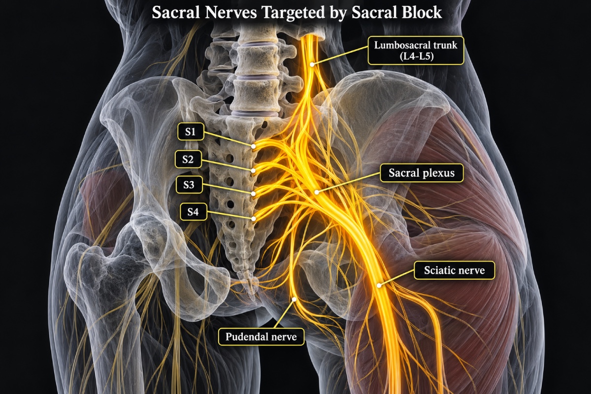
Successful block at the sacral level should be confirmed with several complementary findings. Correct needle placement does not always guarantee adequate anesthesia because local anesthetic may spread unevenly or fail to reach the intended sacral nerve roots. Confirmation should include assessment of needle position, injectate spread, sensory changes, and motor function when appropriate.
Ultrasound can be useful when performing a caudal epidural technique for sacral anesthesia. It allows visualization of structures such as the sacral hiatus, sacral cornua, and sacrococcygeal ligament. Real-time ultrasound may also help identify the needle as it enters the caudal epidural space and can provide evidence of fluid spread during injection (1). Color-flow Doppler may further demonstrate movement of injectate within the epidural space (2). These methods can help reduce uncertainty when surface landmarks are difficult to identify. Negative aspiration for blood or cerebrospinal fluid should also be performed before injection. However, negative aspiration alone does not establish that an effective block has been achieved.
Once sufficient time has passed for the local anesthetic to take effect, sensory examination becomes an important part of confirming successful block at the intended sacral level. Testing should focus on the sacral dermatomes that correspond to the surgical area. The S1 dermatome can be assessed along the lateral aspect of the foot. More caudal sacral segments can be evaluated over the posterior thigh or perineal region when clinically appropriate. Cold sensation and pinprick are commonly used to determine whether sensory transmission has been reduced. Light touch may also be assessed. Different sensory modalities may be affected to different degrees during neuraxial anesthesia (3). For this reason, the clinician should document which method was used and compare the blocked area with an unaffected region.
Both sides of the block should be tested because epidural spread may be asymmetric. A unilateral or patchy loss of sensation may indicate incomplete spread of local anesthetic. Repeated assessment can also be useful because the density and distribution of the block may continue to develop after the initial injection. Testing immediately before the procedure provides additional confirmation that the required sacral segments are adequately anesthetized.
Motor assessment can provide supportive information but should not be used as the primary measure of sacral blockade. Reduced plantar flexion may indicate involvement of the S1 or S2 nerve roots. Weakness of other lower-limb movements may also occur with a dense block. However, substantial sensory anesthesia can occur without marked motor weakness. This is particularly true when lower concentrations of local anesthetic are used. Preserved motor function therefore does not necessarily indicate block failure.
Electrical stimulation can provide a more objective method of evaluating sensory blockade when the standard clinical examination is difficult. Changes in stimulation threshold can reflect reduced sensory transmission as epidural anesthesia develops (4). In routine clinical practice, however, repeated examination with cold, pinprick, or light touch is generally the preferred approach.
Hemodynamic responses should be interpreted cautiously. Stable blood pressure or heart rate does not prove that the sacral dermatomes are adequately blocked. Lack of movement during surgery is also unreliable in a heavily sedated or anesthetized patient. The strongest confirmation of successful sacral block comes from agreement between several findings. Appropriate needle location, expected injectate spread, adequate onset time, and bilateral reduction of sensation within the required sacral dermatomes together provide a more reliable indication of successful sacral blockade.
References
1. Jain D, Hussain SY, Ayub A. Comparative evaluation of landmark technique and ultrasound-guided caudal epidural injection in pediatric population: A systematic review and meta-analysis. Paediatr Anaesth. 2022;32(1):35-42. doi:10.1111/pan.14332
2. Tsui B, Leipoldt C, Desai S. Color flow Doppler ultrasonography can distinguish caudal epidural injection from intrathecal injection. Anesth Analg. 2013;116(6):1376-1379. doi:10.1213/ANE.0b013e31828e5e93
3. Casellato JF, Balki M, Wang A, Ye XY, Downey K, Carvalho JCA. Differential sensory block during labour epidural analgesia: a prospective observational study to investigate the relationship of lower and upper sensory block levels to cold, pinprick, and light touch. Can J Anaesth. 2024;71(6):802-807. doi:10.1007/s12630-023-02638-5
4. Dyhre H, Renck H, Andersson C. Assessment of sensory block in epidural anaesthesia by electric stimulation. Acta Anaesthesiol Scand. 1994;38(6):594-600. doi:10.1111/j.1399-6576.1994.tb03958.x