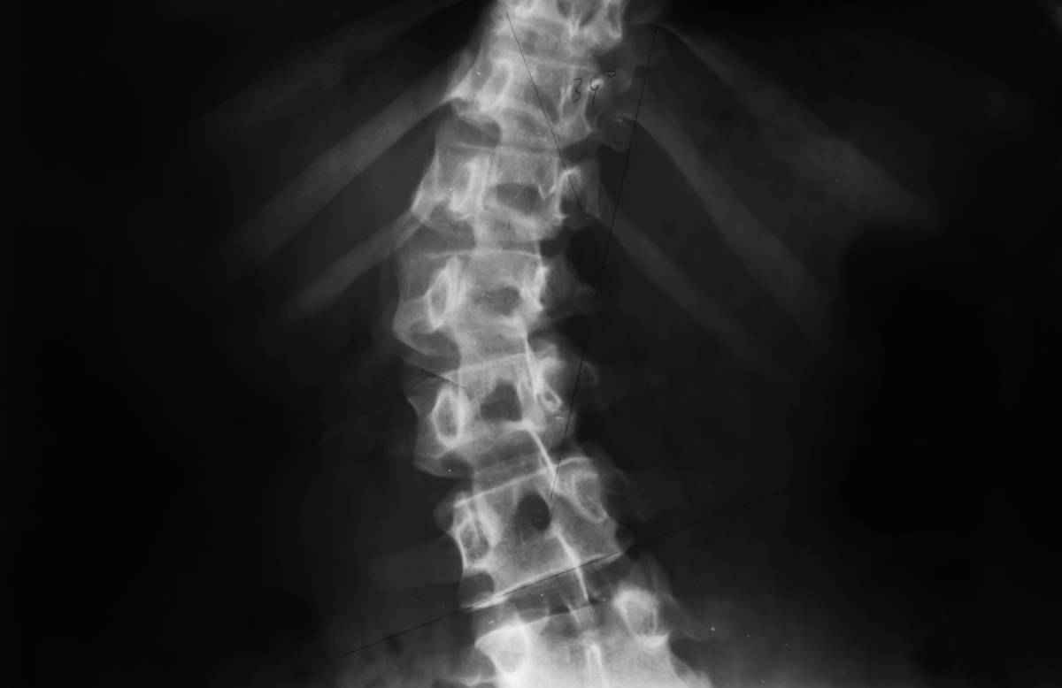
Neuraxial anesthesia is an integral part of perioperative pain management, especially for procedures involving the lower extremities. It involves the administration of anesthetic agents near the spinal cord to block nerve conduction below the level of administration, effectively numbing the lower body. This technique includes spinal anesthesia, epidural anesthesia, and combined spinal-epidural anesthesia, which uses aspects of both. The administration of neuraxial anesthesia in patients with scoliosis is considerably more challenging due to the anatomical distortions present, which can affect both the technical execution of anesthesia and the distribution of the anesthetic agent (1).
Scoliosis, characterized by lateral and/or rotational curvature of the spine, disrupts the regular alignment and positioning of vertebral landmarks that anesthesiologists rely on for safe needle insertion during neuraxial blocks. This disruption can complicate the procedure, increasing the risk of inadequate anesthesia or more serious complications like nerve damage or dural puncture. Therefore, a comprehensive and careful preoperative assessment is critical. This typically includes the use of advanced imaging modalities such as MRI or CT scans which provide detailed views of spinal curvature to assist the anesthesiologist in planning the approach for the neuraxial block approach and anticipating and mitigating procedural challenges (2). Given the irregular anatomical landmarks in patients with scoliosis, anesthesiologists often need to modify traditional techniques. The paramedian approach is often preferred to the standard midline approach because it can more effectively avoid the distorted interspinous processes and provide reliable access to the epidural or subarachnoid space. In addition to adjusting the approach, the use of real-time imaging technologies such as ultrasound or fluoroscopy has become invaluable in guiding needle placement, thereby increasing the accuracy of the procedure and reducing the risks associated with incorrect needle placement (3).
These challenges do not end with needle insertion. The abnormal shape of the spinal canal in scoliosis patients affects the spread of the anesthetic, often resulting in uneven or unpredictable anesthesia. To manage this, anesthesiologists may use techniques such as incremental dosing, in which the anesthetic is administered in controlled, gradual amounts to observe its spread and effectiveness before administering more. Alongside the potential placement of a continuous infusion catheter, this method allows for real-time adjustments to ensure adequate and consistent anesthesia throughout the procedure (4).
Postoperative care is also more complex in patients with scoliosis. The potential for uneven anesthetic spread can result in areas of inadequate pain control, necessitating a more dynamic and responsive postoperative pain management strategy. This often involves a multimodal approach that combines different types of analgesics to effectively manage pain while minimizing side effects (5).
Looking ahead, the field of neuraxial anesthesia in patients with scoliosis will benefit significantly from technological advancements. An innovation like augmented reality could provide anesthesiologists with real-time 3D visual overlays of the patient’s spinal anatomy during the procedure. Additionally, predictive modeling using artificial intelligence may help predict anesthetic spread based on individual spinal geometries. Such exciting developments may further enhance the precision and safety of neuraxial anesthesia administration in this challenging patient population (5).
In summary, neuraxial anesthesia in patients with scoliosis requires a deep understanding of spinal anatomy, precise modification of technique, and vigilant management of anesthetic spread and postoperative pain. Ongoing advancements in technology and technique continue to improve the outcomes for these patients, making it possible to manage their pain effectively and safely during and after surgical procedures.
References
- Goel A, Jeelani TM, Saleem B. Study of spinal anesthesia in patients with scoliosis at a tertiary hospital. Asian J Med Sci. 2021;12(4):345-350.
- Ballarapu GK, Nallam SR, Samantaray A, Kumar VAK, Reddy AP. Thoracolumbar curve and Cobb angle in determining spread of spinal anesthesia in Scoliosis. An observational prospective pilot study. Indian J Anaesth. 2020;64(7):594-598. doi:10.4103/ija.IJA_914_19
- Huang J. Paramedian approach for neuroaxial anesthesia in parturients with scoliosis. Anesth Analg. 2010;111(3):821-822. doi:10.1213/ANE.0b013e3181e6389a
- Ko JY, Leffert LR. Clinical implications of neuraxial anesthesia in the parturient with scoliosis. Anesth Analg. 2009;109(6):1930-1934. doi:10.1213/ANE.0b013e3181bc3584
- Parr WCH, Burnard JL, Wilson PJ, Mobbs RJ. 3D printed anatomical (bio)models in spine surgery: clinical benefits and value to health care providers. J Spine Surg. 2019;5(4):549-560. doi:10.21037/jss.2019.12.07
